Search This Blog

Showing posts with label biology. Show all posts

Wednesday, September 11, 2013

Radiotherapy in Girls and the Risk of Breast Cancer Later in Life

SEPTEMBER 11, 2013
Lynn Yarris (510) 486-5375  lcyarris@lbl.gov
 11 


Science Short
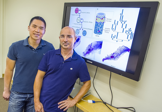
Jonathan Tang (left) and Sylvain Costes developed an agent-based model that is the first multi-scale model of the development of a  mammary gland from the onset of puberty all the way to adulthood. (Photo by Roy Kaltschmidt)
Jonathan Tang (left) and Sylvain Costes developed an agent-based model that is the first multi-scale model of the development of a mammary gland from the onset of puberty all the way to adulthood. (Photo by Roy Kaltschmidt)
Exposing young women and girls under the age of 20 to ionizing radiation can substantially raise the risk of their developing breast cancer later in life. Scientists may now know why. A collaborative study, in which Berkeley Lab researchers played a pivotal role, points to increased stem cell self-renewal and subsequent mammary stem cell enrichment as the culprits. Breasts enriched with mammary stem cells as a result of ionizing irradiation during puberty show a later-in-life propensity for developing ER negative tumors – cells that do not have the estrogen receptor. Estrogen receptors – proteins activated by the estrogen hormone – are critical to the normal development of the breast and other female sexual characteristics during puberty.
“Our results are in agreement with epidemiology studies showing that radiation-induced human breast cancers are more likely to be ER negative than are spontaneous breast cancers,” says Sylvain Costes, a biophysicist with Berkeley Lab’s Life Sciences Division. “This is important because ER negative breast cancers are less differentiated, more aggressive, and often have a poor prognosis compared to the other breast cancer subtypes.”
Costes and Jonathan Tang, also with Berkeley Lab’s Life Sciences Division, were part of a collaboration led by Mary Helen Barcellos-Hoff, formerly with Berkeley Lab and now at the New York University School of Medicine, that investigated the so-called  “window of susceptibility” known to exist between radiation treatments at puberty and breast cancer risk in later adulthood. The key to their success were two mammary lineage agent-based models (ABMs) they developed in which a system is modeled as a collection of autonomous decision-making entities called agents. One ABM simulated the effects of radiation on the mammary gland during either the developmental stages or during adulthood. The other simulated the growth dynamics of human mammary epithelial cells in culture after irradiation.
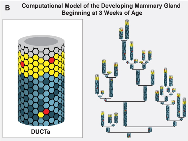
This example from mammary gland agent-based model depicts the network structure at week three formed by cell agents that come together to form a duct agent that in turn organize into a network similar to the branched structure of the mammary gland.
This mammary gland agent-based model depicts the network structure at week three formed by cell agents that come together to form duct agents. In turn, duct agents organize into a network similar to the branched structure of the mammary gland.
“Our mammary gland ABM consisted of millions of agents, with each agent representing either a mammary stem cell, a progenitor cell or a differentiated cell in the breast,” says Tang. “We ran thousands of simulations on Berkeley Lab’s Lawrencium supercomputer during which each agent continually assessed its situation and made decisions on the basis of a set of rules that correspond to known or hypothesized biological properties of mammary cells. The advantage of this approach is that it allows us to view the global consequences to the system that emerge over time from our assumptions about the individual agents. To our knowledge, our mammary gland model is the first multi-scale model of the development of full glands starting from the onset of puberty all the way to adulthood.”
Epidemiological studies have shown that girls under 20 given radiotherapy treatment for disorders such as Hodgkin’s lymphoma run about the same risk of developing breast cancer in their 40s as women who were born with a BRCA gene mutation. From their study, Costes, Tang and their collaboration partners concluded that self-renewal of stem cells was the most likely responsible mechanism.
“Stem cell self-renewal was the only mechanism in the mammary gland model that led to predictions that were consistent with data from both our in vivo mouse work and our in vitro experiments with MCF10A, a human mammary epithelial cell line,” Tang says. “Additionally, our model predicts that this mechanism would only generate more stem cells during puberty while the gland is developing and considerable cell proliferation is taking place.”
Costes and Tang are now looking for genetic or phenotypic biomarkers that would identify young girls who are at the greatest breast cancer risk from radiation therapy. The results of their study with Barcellos-Hoff and her research group show that the links between ionizing radiation and breast cancer extend beyond DNA damage and mutations.
“Essentially, exposure of the breast to ionizing radiation generates an overall biochemical signal that tells the system  something bad happened,” Costes says. “If exposure takes place during puberty, this signal triggers a regenerative response leading to a larger pool of stem cells, thereby increasing the chance of developing aggressive ER negative breast cancers later in life.”
Additional Information
The results of this collaborative study have been published in the journal Stem Cells under the title “Irradiation of juvenile, but not adult, mammary gland increases stem cell self-renewal and estrogen receptor negative tumors.” Tang is the lead author, Barcellos-Hoff is the corresponding author. Other authors in addition to Costes are Ignacio Fernandez-Garcia, Sangeetha Vijayakumar, Haydeliz Martinez-Ruiz, Irineu Illa-Bochaca, David Nguyen and Jian-Hua Mao.

Friday, June 14, 2013

Biological Soil Crust Secrets Uncovered


Berkeley Lab Team Reveals How Desert Cyanobacteria Awaken to Rainfall Then Resume Their Sleep

JUNE 14, 2013
Lynn Yarris (510) 486-5375  lcyarris@lbl.gov
 14 


Feature
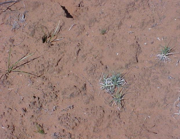
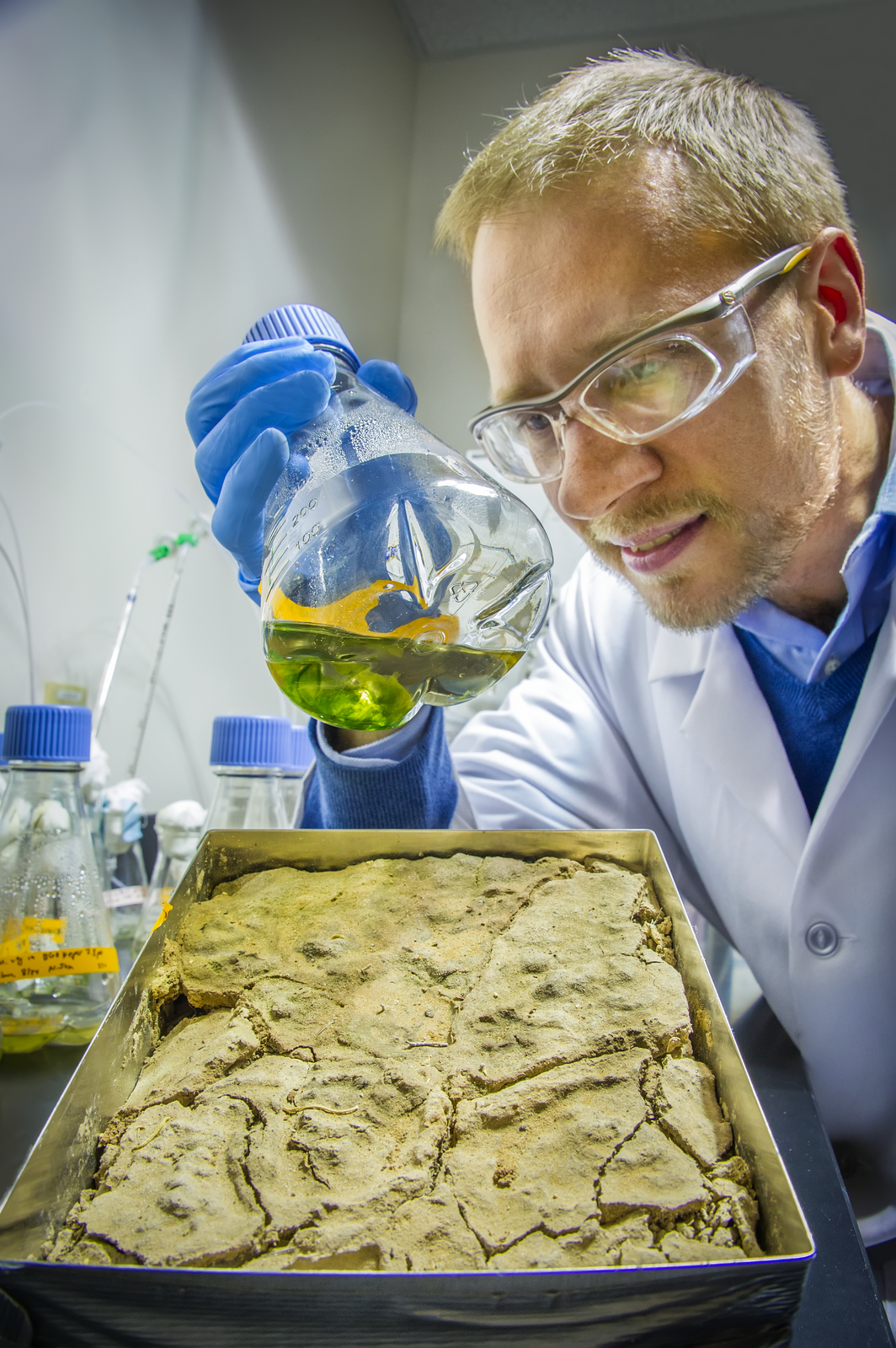
This looks like ordinary dirt but it is a biological soil crust (BSC), a living mantle of soil particles bound together by microbes and their by-products. BSCs are common to the arid and semiarid lands that make up about 40-percent of Earth’s total land mass. (Photo from Ferran Garcia-Pichel)
They lie dormant for years, but at the first sign of favorable conditions they awaken. This sounds like the tagline for a science fiction movie, but it describes the amazing life-cycles of microbial organisms that form the biological soil crusts (BSCs) of Earth’s deserts. Now a research team with the U.S. Department of Energy (DOE)’s Lawrence Berkeley National Laboratory (Berkeley Lab) has reported a unique molecular-level analysis of a BSC cyanobacterium responding to the wetting and drying of its environment. The results hold implications for land management, improved climate change models, and a better understanding of carbon cycling in soil microbial communities and how changes in global temperatures impact Earth’s deserts.
“We found a way to measure from start to finish in real unaltered samples the molecular events behind the response of cyanobacterium to wetting and drying in a desert BSC,” says Aindrila Mukhopadhyay, a biologist with Berkeley Lab’s Physical Biosciences Division. “Not only did we get a good view of the genetic machinery that wakes the microbes up, but we also got a good sense of what constitutes a healthy BSC.”
Mukhopadhyay and Trent Northen, a chemist with Berkeley Lab’s Life Sciences Division, are the corresponding authors of a paper describing this research in the journal of the International Society for Microbial Ecology. The paper is titled “Dynamic cyanobacterial response to hydration and dehydration in a desert biological soil crust.”
Arid and semi-arid deserts make up about 40-percent of Earth’s total land mass. Much of the undisturbed soil crust in these deserts is a living mantle of microbes and their by-products, with the predominant inhabitants being cyanobacteria, microorganisms that use photosynthesis for energy. To survive dry spells that can go on for years, BSC microorganisms must enter a dormant state but they must also be poised for rapid resuscitation to utilize short periods of precipitation. For a better understanding of how the microbes are able to do this, the Berkeley Lab research team studied the cyanobacteriumMicrocoleus vaginatus.
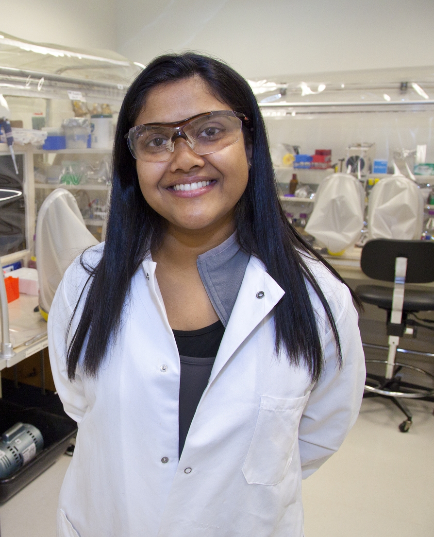
Aindrila Mukhopadhyay is a biologist with Berkeley Lab’s Physical Biosciences Division (Photo by Roy Kaltschmidt)
“M. vaginatus is the early BSC colonizer, an ecosystem pioneer that fixes carbon and binds the soil together allowing the community to develop while preventing wind erosion,” says Northen. “It is globally distributed in regions where water is scarce, including not only hot deserts but the arctic as well.”
Working with the research group of Ferran Garcia-Pichel at Arizona State University, the Berkeley Lab team collected pie-sized samples of soil crust from the cold desert area near Moab, Utah. These samples were subjected to in situwetting and drying over a period of six days, including two diel (day/night) cycles. The wetting simulated conditions that would be brought about by the passage of a rain front. During the study, the researchers performed whole-genome transcriptional analysis and precise biochemical measurements.
“Simulated weather conditions and sampling in a temporal manner over the six day experimental period allowed us to track key transcriptional and metabolic responses as they occurred,” says Mukhopadhyay. “We found that within three minutes after wetting began, metabolic processes in the dormant microbial cells came alive. Within one hour, photosynthesis began, accompanied by carbon dioxide up-take. During this time we saw specific genes get turned on/off and specific gene expressions be elevated or depressed.”
The rain episode lasted three days. Although rare, such conditions do occur in the desert and enabled the researchers to differentiate between wetting-up and drying-down responses from diel cycle responses. They discovered that when dehydration of the soil resumed, the mechanisms triggered by wetting were mirrored in the reverse direction, suggesting that the M. vaginatus microbes were preparing themselves to re-enter desiccation-induced dormancy.
“These BSC microbes are highly attuned to their environment and they respond very quickly to changes,” Mukhopadhyay says. “Their responses, however, are highly sensitive to physical disturbances and alterations in temperature.”
In their paper, the Berkeley Lab researchers say that a better understanding of how environmental factors influence the growth and metabolism of M. vaginatus and other BSC microbes should improve our ability to predict the impacts of climate or land-use change on BSC systems. This knowledge should also help in the development of management strategies for restoring BSCs following large-scale disturbances.
Trent Northen is a chemist with Berkeley Lab’s Life Sciences Division (Photo by Roy Kaltschmidt)
“BSC systems represent the world’s largest biofilms, and the demise of such systems could release carbon, and probably more importantly, dust into the atmosphere, changing the albedo of snow-packs,” says Northen. “Restoring or protecting BSCs through changes in land-use could help pull carbon out of the atmosphere and reduce dust.”
A better understanding of BSCs could also deepen our understanding of the carbon cycle, which could improve the accuracy of climate models, as Mukhopadhyay explains.
“In most climate models there is little or no accounting for the carbon fixed by soil microbes,” she says. “BSC cyanobacteria are the photosynthetic organism for deserts and understanding their role in the carbon-cycle will help fill in current climate model gaps. This should help improve the accuracy of these models.”
The Berkeley Lab team would like to expand their efforts to examine other BSC systems around the world. Among other issues, they would like to identify the signaling mechanisms that inform dormant BSC microbes that moisture is present or that the temperature is changing and it is time to respond.
“BSCs are sentinels for marking the health or decline of the world’s deserts, one of Earth’s major ecosystems,” Mukhopadhyay says. “It is vital that we understand them.”
Co-authoring the paper “Dynamic cyanobacterial response to hydration and dehydration in a desert biological soil crust” were Lara Rajeev, Ulisses Nunes da Rocha, Niels Klitgord, Eric Luning, Julian Fortney, Seth Axen, Patrick Shih, Nicholas Bouskill, Benjamin Bowen, Cheryl Kerfeld, Ferran Garcia-Pichel and Eoin Brodie.
This research was funded through Berkeley Lab’s Laboratory Directed Research and Development program.
#  #  #
Lawrence Berkeley National Laboratory addresses the world’s most urgent scientific challenges by advancing sustainable energy, protecting human health, creating new materials, and revealing the origin and fate of the universe. Founded in 1931, Berkeley Lab’s scientific expertise has been recognized with 13 Nobel prizes. The University of California manages Berkeley Lab for the U.S. Department of Energy’s Office of Science. For more, visit www.lbl.gov.
DOE’s Office of Science is the single largest supporter of basic research in the physical sciences in the United States, and is working to address some of the most pressing challenges of our time. For more information, please visit the Office of Science website atscience.energy.gov/.
Additional Information
A video of Aindrila Mukhopadhyay discussing aspects of her BSC research can be viewedhere

Monday, June 3, 2013

Berkeley Lab Researchers Discover How and Where Breast Tumor Cells Become Dormant and What Causes Them to Become Metastatic

News Release:


JUNE 03, 2013
Lynn Yarris (510) 486-5375  lcyarris@lbl.gov
 7 


News Release
A new study shows that stable microvasculature constitutes a dormant niche for disseminated breast cancer cells, whereas a sprouting neovasculature (green points) promotes breast tumor cell growth.
The long-standing mystery behind dormant disseminated breast tumor cells and what activates them after years and even decades of latency may have been solved. Researchers with the U.S. Department of Energy (DOE)’s Lawrence Berkeley National Laboratory (Berkeley Lab) have identified the microenvironment surrounding microvasculature – the small blood vessels that transport blood within tissues – as a niche where dormant cancer cells reside. When these blood vessels begin to sprout, the new tips produce molecules that transform dormant cancer cells into metastatic tumors.
In a small but significant number of breast cancer patients, cancerous cells can move through the bloodstream from breast tissue to secondary sites in other parts of the body where they may remain in a dormant state, clinically undetected, for an extended period of time, before suddenly becoming metastatic. It has been difficult if not impossible to predict if and when metastases will occur.
“Our study reveals that a stable microvasculature constitutes a dormant niche, whereas a sprouting neovasculature sparks micrometastatic outgrowth,” says cell biologist Mina Bissell, in whose laboratory this work was done. “Sprouting is meant to coincide with tissue growth, but if a tumor cell happens to be in the wrong place at the wrong time, then it comes under the influence of the factors deposited by tip cells and it starts growing.”
Bissell, Distinguished Scientist with Berkeley Lab’s Life Sciences Division ranks among the world’s foremost breast cancer researchers. She is one of two corresponding authors – the other is Cyrus Ghajar a bioengineer and member of Bissell’s research group – of a paper describing this study in the journal Nature Cell Biology. The paper is titled “The perivascular niche regulates breast tumor dormancy.”
“Some patients may experience metastatic relapse within months, other patients may go several years or even decades without distant recurrence,” Bissell says. “The recent discovery of tumor-promoting milieus, referred to as metastatic niches, that are established at distant sites prior to or upon the arrival of disseminated tumor cells could explain cancer cells that relapse early, but in late relapsing populations, what tumor cells do from the time of dissemination to the time they become clinically detectable has been a big question.”
Mina Bissell and Cyrus Ghajar may have solved the long-standing mystery behind dormant disseminated tumor cells and what activates them after years and even decades of latency. (Photo by Roy Kaltschmidt)
Previous research by Bissell and her group showed that the basement membrane, the thin layer of extracellular matrix proteins surrounding breast and other epithelial tissue, provides a microenvironment that induces quiescence in normal epithelial cells and dormant tumor cells alike. Given that breast cancer cells traveling through the bloodstream on their way to secondary sites where breast tumors metastasize most often – lung, bone marrow, brain and liver – must first pass through the basement membrane microvasculature, Ghajar and Bissell suspected that the basement membrane could be a major component of the dormant niche in distant organs.
To test this idea, the researchers utilized two mouse models of human breast cancer metastasis and found dormant disseminated tumor cells residing upon the membrane microvasculature of lung, bone marrow and brain tissue. To determine whether endothelial cells – the cells that line the interior surface of blood vessels – directly influence breast cancer cell growth, they then created unique organotypic models of lung and bone marrow microvascular niches, in which endothelial cells formed blood vessel-like structures in culture as they would in the original organ. When tumor cells were placed on top of these blood vessel-like structures, the in vivo observations of the researchers was reproduced.
With their organotypic models, Ghajar, Bissell and their collaborators discovered that the protein thrombospondin-1, which is prevalent in stable microvasculature, creates a dormant niche by suppressing the growth of breast cancer cells. However, when the tips of these blood cells begin to sprout, the thrombospondin-1 proteins give way to TGF-beta 1 and periostin proteins in the neovasculature, turning it into a metastatic niche that not only permits but accelerates the growth of breast cancer cells.
“Ours is the first study to define the dormant niche on a cellular and molecular basis, and it is interesting that the culprit is the tissue we so often assume is a passive bystander, the microvascular endothelium,” Ghajar says. “Moreover, we show that, as with most if not all biological processes, homeostasis is key. In this case, disruption of stable vascular endothelium disrupts this dormant niche and encourages the formation of a microenvironment that sparks micrometastatic outgrowth.”
Ghajar says their findings were a surprise in that “Not only was there a loss of growth suppression at neovascular tips, but there was also a gain of tumor promotion. It was never known that endothelial tip cells have such unique secretion or deposition profiles, let alone shown that these profiles actually favor tumor growth.”
The identification of dormant niches in basement membrane microvasculature and how those niches become metastatic in the neovasculature holds important implications for future breast cancer therapies.
“Our findings point the way to therapies aimed at managing metastatic disease before it even occurs,” Ghajar says. “Dormant disseminated tumors can be ticking time bombs, but now that we know some of the triggers, it may be possible to develop therapies to ensure that disseminated cancer cells remain in a dormant state, or other therapies that eradicate these cells before they form  full-blown metastases.”
The unique organotypic models developed for this study also hold promise for future therapy research.
“Our organotypic models demonstrate the power of the  microenvironment by showing that normal cells in their native architecture naturally steer fully malignant, genotypically aberrant tumor cells into a quiescent state,” Ghajar says.
“In the future, our models should provide robust tools to screen for therapies that impact tumor dormancy and metastasis, and should also provide a platform to solve other biological mysteries that underlie dormancy.”
Ghajar, Bissell and their collaborators are now investigating whether these results with breast cancer tumors also apply to other types of tumors and in other secondary tissues. They are also looking to identify unique mechanisms specific to other tissues that also result in tumor dormancy.
In addition to Bissell and Ghajar, other co-authors of the Nature Cell Biology were Héctor Peinado, Hidetoshi Mori, Irina Matei, Kimberley Evason, Hélène Brazier, Dena Almeida, Antonius Koller, Katherine Hajjar, Didier Stainier, Emily Chen and David Lyden, who is with the Weill Cornell Medical College.
This research was supported in part by the National Cancer Institute and the U.S. Department of Defense.
# # #
Lawrence Berkeley National Laboratory addresses the world’s most urgent scientific challenges by advancing sustainable energy, protecting human health, creating new materials, and revealing the origin and fate of the universe. Founded in 1931, Berkeley Lab’s scientific expertise has been recognized with 13 Nobel prizes. The University of California manages Berkeley Lab for the U.S. Department of Energy’s Office of Science. For more, visit www.lbl.gov.

Additional Information
For more on Mina Bissell’s breast cancer research go here