News Release

A new study shows that stable microvasculature constitutes a dormant niche for disseminated breast cancer cells, whereas a sprouting neovasculature (green points) promotes breast tumor cell growth.
The long-standing mystery behind dormant disseminated breast tumor cells and what activates them after years and even decades of latency may have been solved. Researchers with the U.S. Department of Energy (DOE)’s Lawrence Berkeley National Laboratory (Berkeley Lab) have identified the microenvironment surrounding microvasculature – the small blood vessels that transport blood within tissues – as a niche where dormant cancer cells reside. When these blood vessels begin to sprout, the new tips produce molecules that transform dormant cancer cells into metastatic tumors.
In a small but significant number of breast cancer patients, cancerous cells can move through the bloodstream from breast tissue to secondary sites in other parts of the body where they may remain in a dormant state, clinically undetected, for an extended period of time, before suddenly becoming metastatic. It has been difficult if not impossible to predict if and when metastases will occur.
“Our study reveals that a stable microvasculature constitutes a dormant niche, whereas a sprouting neovasculature sparks micrometastatic outgrowth,” says cell biologist Mina Bissell, in whose laboratory this work was done. “Sprouting is meant to coincide with tissue growth, but if a tumor cell happens to be in the wrong place at the wrong time, then it comes under the influence of the factors deposited by tip cells and it starts growing.”
Bissell, Distinguished Scientist with Berkeley Lab’s Life Sciences Division ranks among the world’s foremost breast cancer researchers. She is one of two corresponding authors – the other is Cyrus Ghajar a bioengineer and member of Bissell’s research group – of a paper describing this study in the journal
Nature Cell Biology. The paper is titled “The perivascular niche regulates breast tumor dormancy.”
“Some patients may experience metastatic relapse within months, other patients may go several years or even decades without distant recurrence,” Bissell says. “The recent discovery of tumor-promoting milieus, referred to as metastatic niches, that are established at distant sites prior to or upon the arrival of disseminated tumor cells could explain cancer cells that relapse early, but in late relapsing populations, what tumor cells do from the time of dissemination to the time they become clinically detectable has been a big question.”

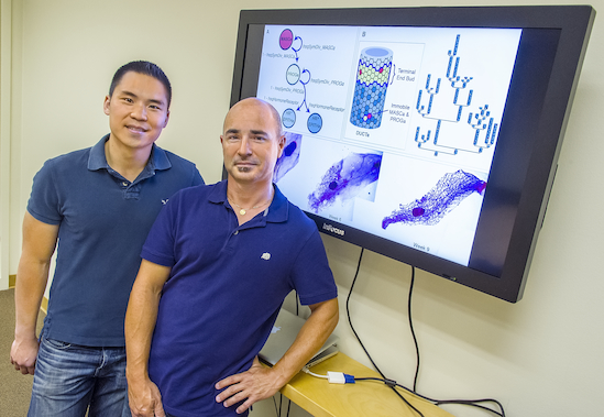
Mina Bissell and Cyrus Ghajar may have solved the long-standing mystery behind dormant disseminated tumor cells and what activates them after years and even decades of latency. (Photo by Roy Kaltschmidt)
Previous research by Bissell and her group showed that the basement membrane, the thin layer of extracellular matrix proteins surrounding breast and other epithelial tissue, provides a microenvironment that induces quiescence in normal epithelial cells and dormant tumor cells alike. Given that breast cancer cells traveling through the bloodstream on their way to secondary sites where breast tumors metastasize most often – lung, bone marrow, brain and liver – must first pass through the basement membrane microvasculature, Ghajar and Bissell suspected that the basement membrane could be a major component of the dormant niche in distant organs.
To test this idea, the researchers utilized two mouse models of human breast cancer metastasis and found dormant disseminated tumor cells residing upon the membrane microvasculature of lung, bone marrow and brain tissue. To determine whether endothelial cells – the cells that line the interior surface of blood vessels – directly influence breast cancer cell growth, they then created unique organotypic models of lung and bone marrow microvascular niches, in which endothelial cells formed blood vessel-like structures in culture as they would in the original organ. When tumor cells were placed on top of these blood vessel-like structures, the
in vivo observations of the researchers was reproduced.
With their organotypic models, Ghajar, Bissell and their collaborators discovered that the protein thrombospondin-1, which is prevalent in stable microvasculature, creates a dormant niche by suppressing the growth of breast cancer cells. However, when the tips of these blood cells begin to sprout, the thrombospondin-1 proteins give way to TGF-beta 1 and periostin proteins in the neovasculature, turning it into a metastatic niche that not only permits but accelerates the growth of breast cancer cells.
“Ours is the first study to define the dormant niche on a cellular and molecular basis, and it is interesting that the culprit is the tissue we so often assume is a passive bystander, the microvascular endothelium,” Ghajar says. “Moreover, we show that, as with most if not all biological processes, homeostasis is key. In this case, disruption of stable vascular endothelium disrupts this dormant niche and encourages the formation of a microenvironment that sparks micrometastatic outgrowth.”
Ghajar says their findings were a surprise in that “Not only was there a loss of growth suppression at neovascular tips, but there was also a gain of tumor promotion. It was never known that endothelial tip cells have such unique secretion or deposition profiles, let alone shown that these profiles actually favor tumor growth.”
The identification of dormant niches in basement membrane microvasculature and how those niches become metastatic in the neovasculature holds important implications for future breast cancer therapies.
“Our findings point the way to therapies aimed at managing metastatic disease before it even occurs,” Ghajar says. “Dormant disseminated tumors can be ticking time bombs, but now that we know some of the triggers, it may be possible to develop therapies to ensure that disseminated cancer cells remain in a dormant state, or other therapies that eradicate these cells before they form full-blown metastases.”
The unique organotypic models developed for this study also hold promise for future therapy research.
“Our organotypic models demonstrate the power of the microenvironment by showing that normal cells in their native architecture naturally steer fully malignant, genotypically aberrant tumor cells into a quiescent state,” Ghajar says.
“In the future, our models should provide robust tools to screen for therapies that impact tumor dormancy and metastasis, and should also provide a platform to solve other biological mysteries that underlie dormancy.”
Ghajar, Bissell and their collaborators are now investigating whether these results with breast cancer tumors also apply to other types of tumors and in other secondary tissues. They are also looking to identify unique mechanisms specific to other tissues that also result in tumor dormancy.
In addition to Bissell and Ghajar, other co-authors of the
Nature Cell Biology were Héctor Peinado, Hidetoshi Mori, Irina Matei, Kimberley Evason, Hélène Brazier, Dena Almeida, Antonius Koller, Katherine Hajjar, Didier Stainier, Emily Chen and David Lyden, who is with the Weill Cornell Medical College.
This research was supported in part by the National Cancer Institute and the U.S. Department of Defense.
# # #
Lawrence Berkeley National Laboratory addresses the world’s most urgent scientific challenges by advancing sustainable energy, protecting human health, creating new materials, and revealing the origin and fate of the universe. Founded in 1931, Berkeley Lab’s scientific expertise has been recognized with 13 Nobel prizes. The University of California manages Berkeley Lab for the U.S. Department of Energy’s Office of Science. For more, visit
www.lbl.gov.
Additional Information
For more on Mina Bissell’s breast cancer research go
here